The Mystery of Electrical Data Revealed
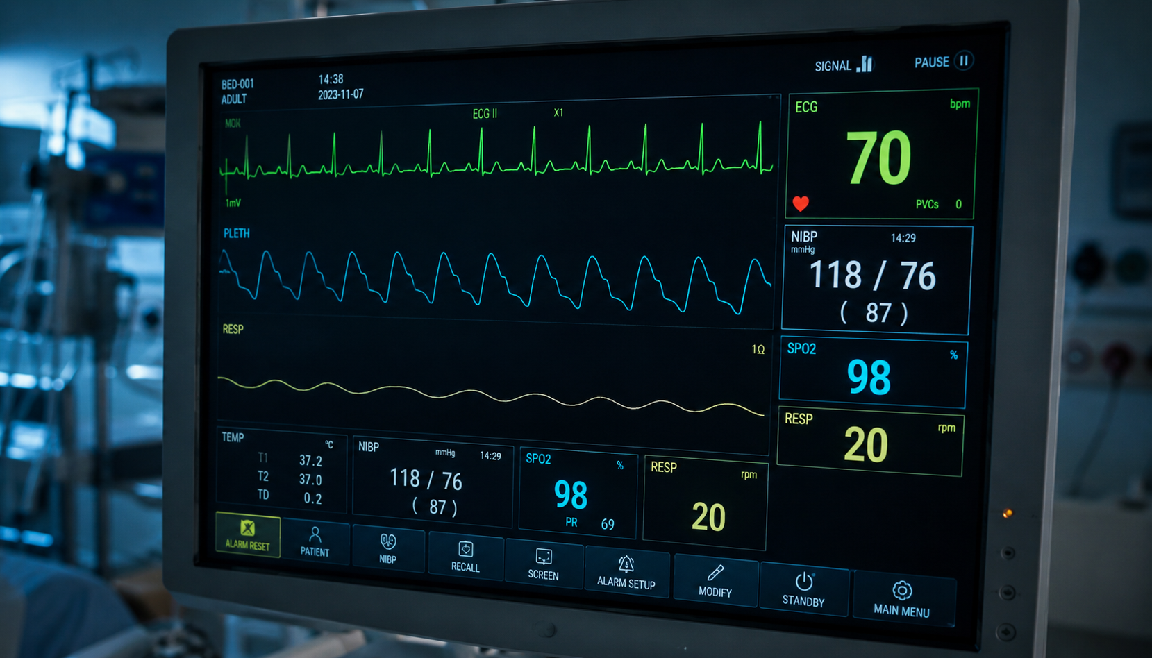
In every ambulance, defibrillators analyze a patient’s heart’s electrical signal before delivering a shock. Historically, the seconds of recording preceding the shock were viewed by doctors as mere background noise—a chaotic jumble on a screen indicating that the heart had stopped, without providing any additional information.
By examining the electrical activity on the surface electrocardiogram (ECG) just before the first defibrillation shock, the researchers found that this data could provide valuable clues. These structural differences in the waveform could genuinely help predict victims’ chances of recovery.
Uneven Resistance to Oxygen Deprivation
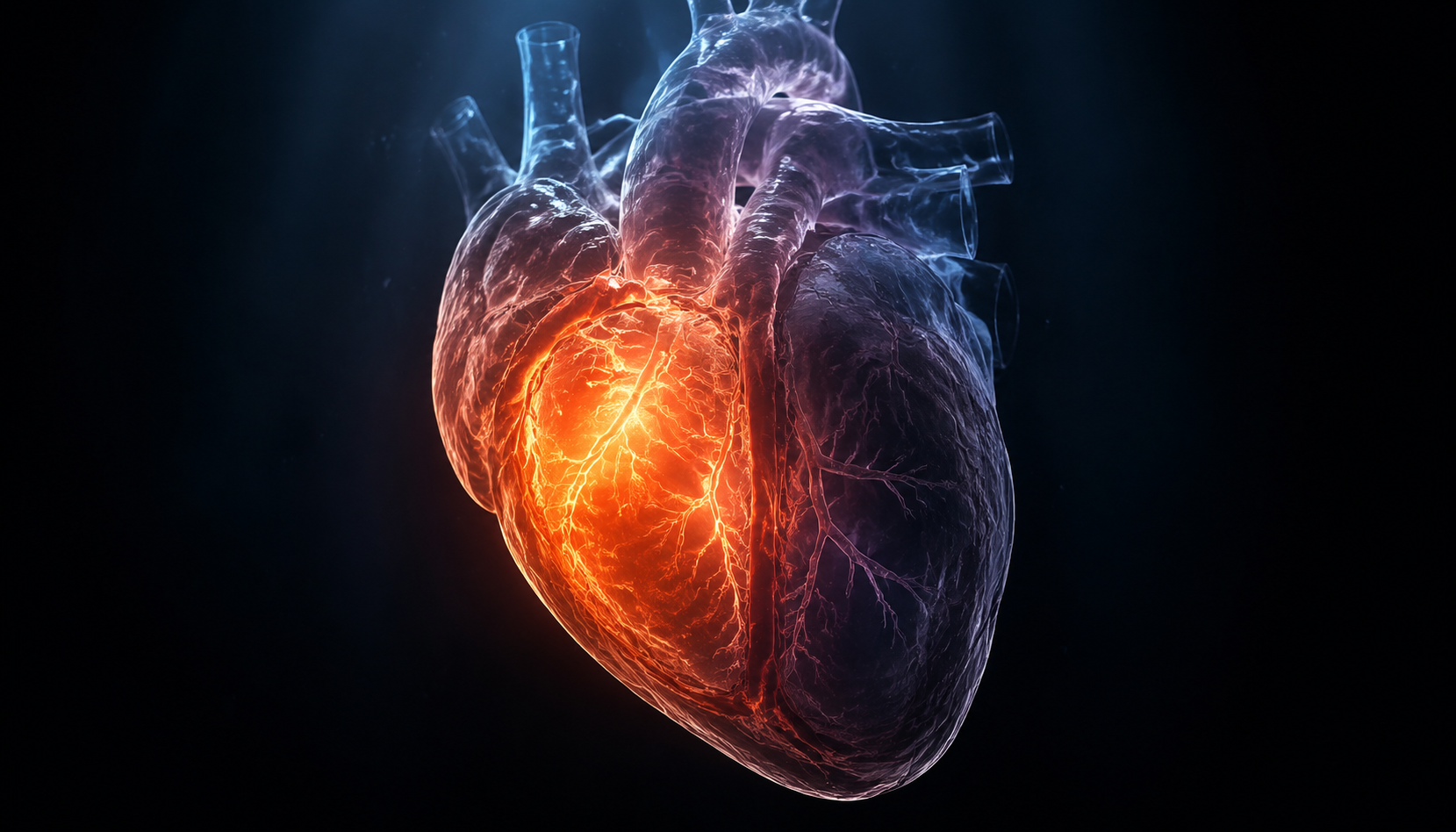
When fibrillation occurs, blood flow ceases within a matter of seconds, plunging the body into a state of collapse known as cardiac arrest. Blood supply is cut off, depriving cells of their essential fuel. Faced with this lack of oxygen—a condition called ischemia—the two lower chambers do not respond at the same rate. The study reveals that the right ventricle tolerates this deprivation significantly better than the left ventricle.
Cross-validation between animal and computational models

To understand in detail what happens inside a failing heart during these frantic minutes, a team from the National Center for Cardiovascular Research (CNIC) in Spain conducted in-depth investigations. Led by Dr. David Filgueiras-Rama, head of the institute’s arrhythmia mechanisms group, the scientists monitored electrical activity second by second.
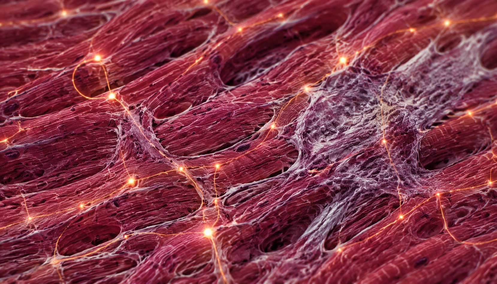
The researchers relied on observations of more than 70 pigs, a species whose heart is very similar in size and structure to ours. To visualize the phenomenon of asymmetric decay, they used a specific technique that converts electrical signals from the heart’s surface into visible light. Under the cameras’ gaze, the left ventricle darkened first, while the right side continued to flicker.
Wire electrodes inserted into each chamber confirmed that the right side fired more quickly. Computer models, developed using measurements specific to the animals, validated this dynamic. Under conditions of a starved heart, the simulated right ventricle retained its excitability—its ability to contract—while the left ventricle lost it earlier, proving unequivocally that electrical silence sets in first on the left.
The initial state of the muscle does not alter this asymmetry
One of the study’s key findings is that this advantage of the right ventricle appears to be deeply embedded within the muscle itself. This pattern was consistently observed in every animal tested, appearing particularly clearly between the inner and outer walls of the heart tissue. The team thus demonstrated that this was not an isolated anomaly in a single experiment.
The right side’s superiority persisted regardless of the heart’s initial health status. Some animals had healthy muscle, while others bore the scars of a controlled heart attack induced weeks earlier. Previous research has linked this type of scar tissue to a significantly higher risk of fibrillation, but the right ventricle’s reserve capacity was not dependent on this underlying condition.
A New Predictive Tool for Emergency Responders
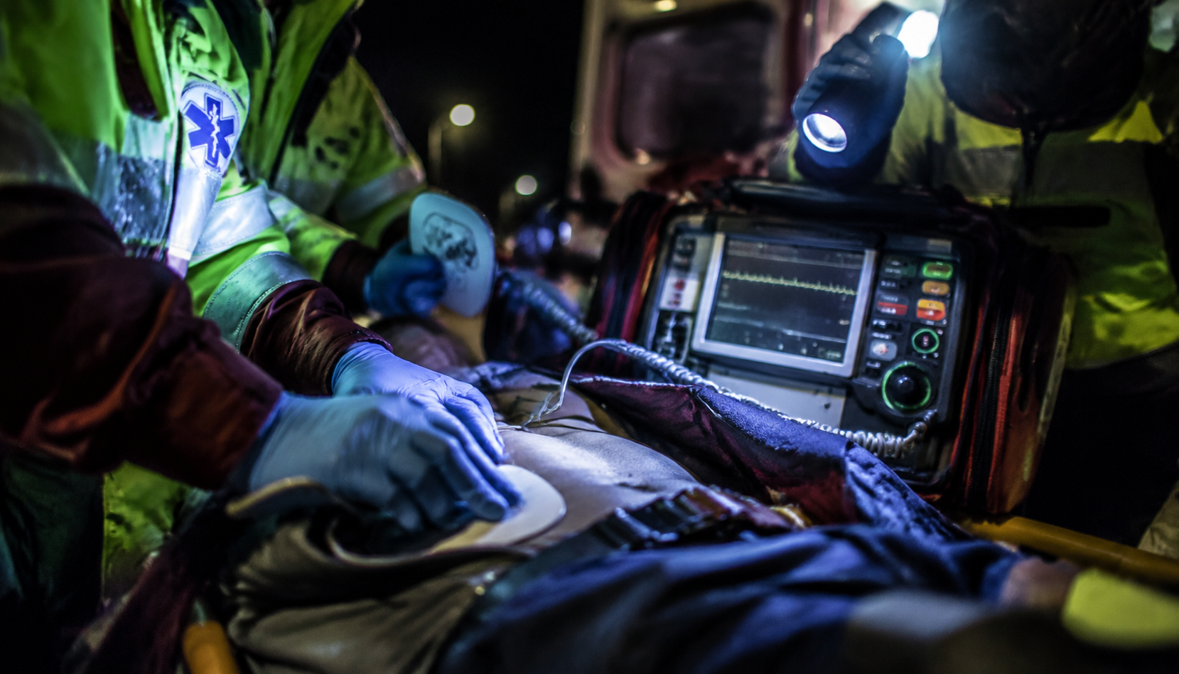
Applying these findings to humans revealed a previously unknown link between residual electrical activity and neurological prognosis. Patients whose hearts were still emitting rapid signals just before the first shock was administered tended to recover with their brains intact. The rhythm of the signal appears to reflect how well the brain itself withstood the lack of blood flow. As the data put it: “A faster trace, a better outlook.”
Predicting damage in a resuscitated patient is one of the most difficult medical decisions to make—a central topic addressed in numerous clinical analyses. Never before has a simple chest recording taken in those first few minutes been so directly linked to the final outcome. This signal, already recorded by every defibrillator in the field, holds the promise of helping emergency teams quickly identify patients with the highest probability of full recovery.
This research establishes that the right ventricle is more resilient during a cardiac event, identifying the left side as the weak point that needs to be strengthened. “The benefit could lie in new therapies aimed at protecting the left ventricle,” said Dr. Jorge García Quintanilla, principal investigator of the research team. For any medical questions, consult a qualified healthcare professional.
Source: earth.com
Cardiac arrest: The right side of the heart keeps functioning longer—a crucial finding for determining prognosis